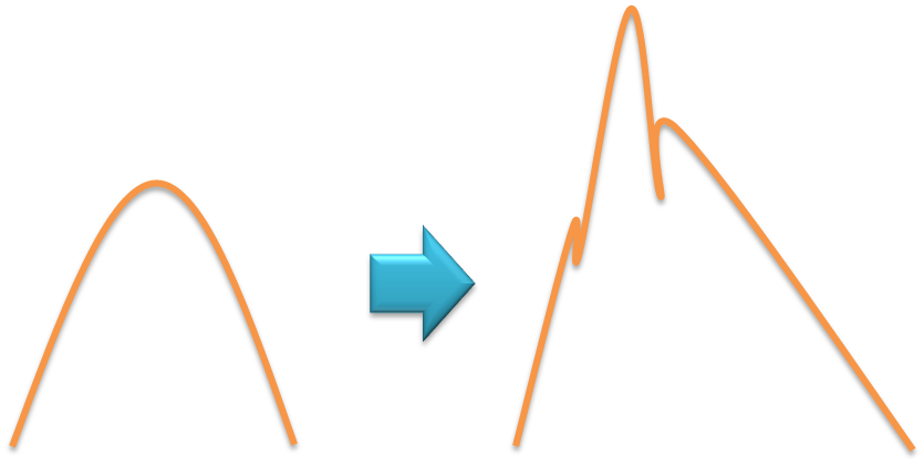
최근 HSCCC를 이용한 AT의 분리에 있어서 단일 성분으로 예상됨에도 불구하고 큰 피크의 모양이 비 대칭이고 조그마한 다른 피크 ( valley peak )가 보여서 왜 그런가 하고 찾아보았다..
Sometime, the ELSD detector can give the peak shape like that, depending on the flow rate and droplets size distribution. If the nebulizer doesn't function well, some large droplets travel later than most fine droplets and it gives additional peak due to higher scattering response. It could be fixed through increasing the gas flow..
ELSD detectors must be optimized for a given flow rate, solvent composition, nebulizer gas flow rate and drift tube temperature (and if a gradient is used, then this is more complex as you must find a compromise in settings). An idea balance between these parameters must be found for each method used. Too high a flow rate relative to the drift tube temperature and/or gas flow rate will cause poor nebulization or even droplets to form which will result in strange signals. In my experience, "split" peaks are usually caused by a clogged or partially plugged nebulizer "spitting" sample out. It can also occur when too high a sample concentration has been injected.
ELSD units require a great deal of maintenance, understanding and optimization time to use. They should never be used like a "plug and play" UV detector which is far easier to use, maintain and clean. That said, ELSD's are useful for sample types where nothing else works well (i.e. some lipids, fats, triglycerides, sugars, polymers...). They key to being successful with an ELSD is spending the time to learn how to use it properly (many test runs to optimize the many parameters stated before as well as regular cleaning). Last Point: They are NOT Universal Detectors (that is an old advertising/marketing phrase only and unscientific).
Source from..
: Chromatography forum